KEY TAKEAWAYS
Noris Medical Pteryfit™ implants are specifically designed to engage to the pterygomaxillary complex and allow for immediately loading protocols.
Noris Medical Zygomatic implants have a unique design especially suited for extra-sinus surgical approaches. The aggressive rough surfaced end cutting tip and smooth mid-coronal shaft make this implant an ideal fixture for treating cases with severe maxillary bone loss.
The Noris Medical line of dental implants are particularly suited for treatment of cases with severe bone loss. Furthermore, with 0, 17, 30, 45, 52, and 60 degree abutments, Noris Medical has one of the widest selection of prosthetic platforms in the industry.
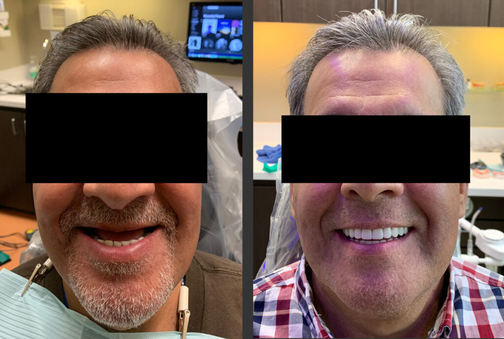
1. Patient before treatment. The patient had been wearing an upper denture and bone atrophy was significant. The mandible had many missing, ailing, and failing teeth.
2. Patient after treatment with zygomatic, pterygoid, vomer, and standard dental implants.
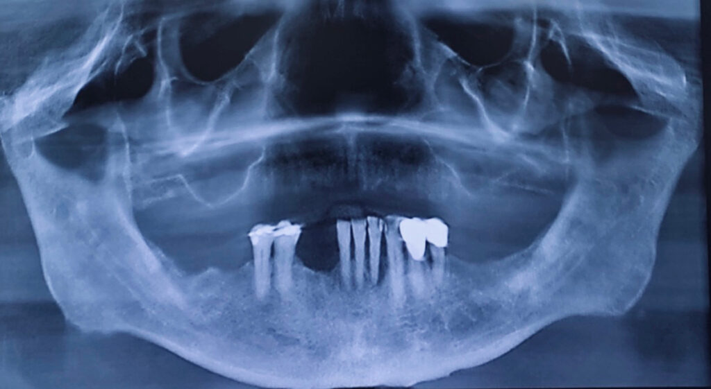
3. Pre-treatment panoramic radiograph showing pneumatized maxillary sinuses, failing mandibular teeth, etc.
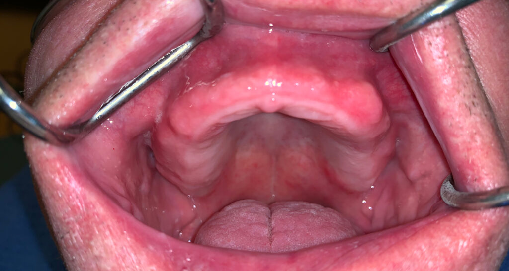
4. Intraoral condition of maxilla before surgery.
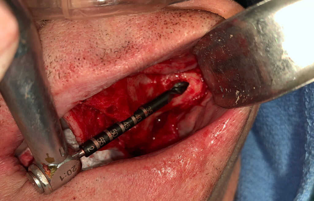
5. Extra-sinus drilling technique for the placement of Noris Medical® zygomatic implants.
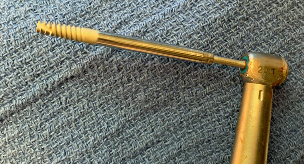
6. Noris Medical® zygomatic implant. (45mm).
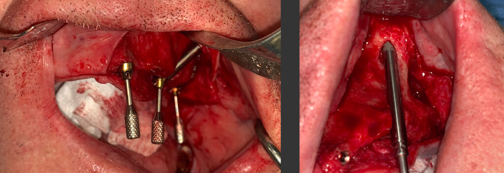
7. Placement of Noris Medical® zygomatic implant using the extra-sinus approach. 8. Noris Medical® Tuff™ implants, zygomatic implants, and Pteryfit™ implants with 17º, 30º, and 45º abutments in place. Note the parallelism achieved due to the wide abutment selection.
9. Maxilla sutured after surgery using Dr. Holtzclaw’s famous “Texas 2-Step” suture protocol.
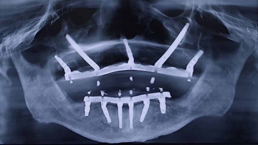
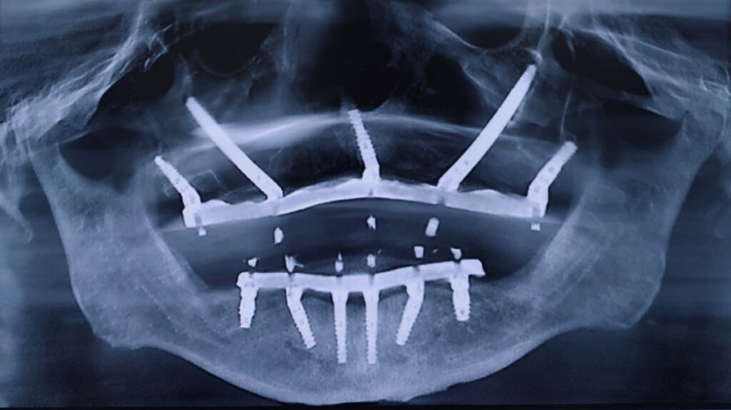
10. Final radiograph after treatment. Maxilla was restored with Noris Medical® Tuff™ implants, zygomatic implants, and Pteryfit™ implants while the mandible was restored with Noris Medical® Tuff™ implants.
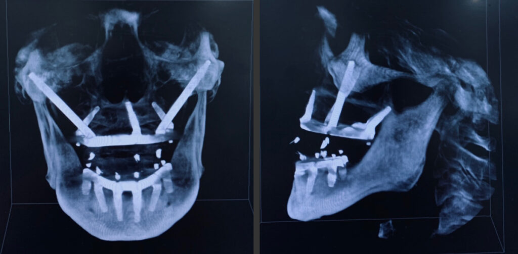
11. CBCT scan (cranial view )of restored case with zygomatic, pterygoid, vomer, and standard dental implants.
12. Lateral CBCT scan of restored case with zygomatic, pterygoid, vomer, and standard dental implants.
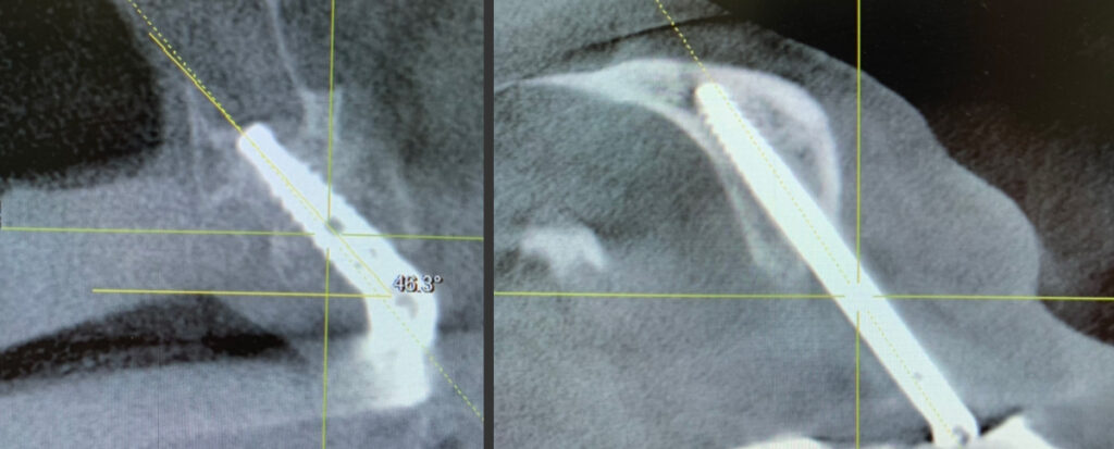
13. CBCT scan of Noris Medical® Pteryfit™ implant engaging the pterygomaxillary complex.

14. CBCT scan of Noris Medical® zygomatic implant engaging the zygoma.